IB Research: A new adenomyosis ultrasound marker
Adenomyosis is a condition that is difficult to diagnose and it is far more common than it is often thought to be. Diagnosis needs to be based on clinical data and confirmed using imaging techniques. Up until now, MRI scans were the go-to technique for providing these necessary images, but ultrasounds may now replace them as a more economically viable and reliable technique. The JZ, or the endometrium-myometrium junctional zone, may be accessed and even measured using a 3D ultrasound and this is particularly true when making use of coronal planes.
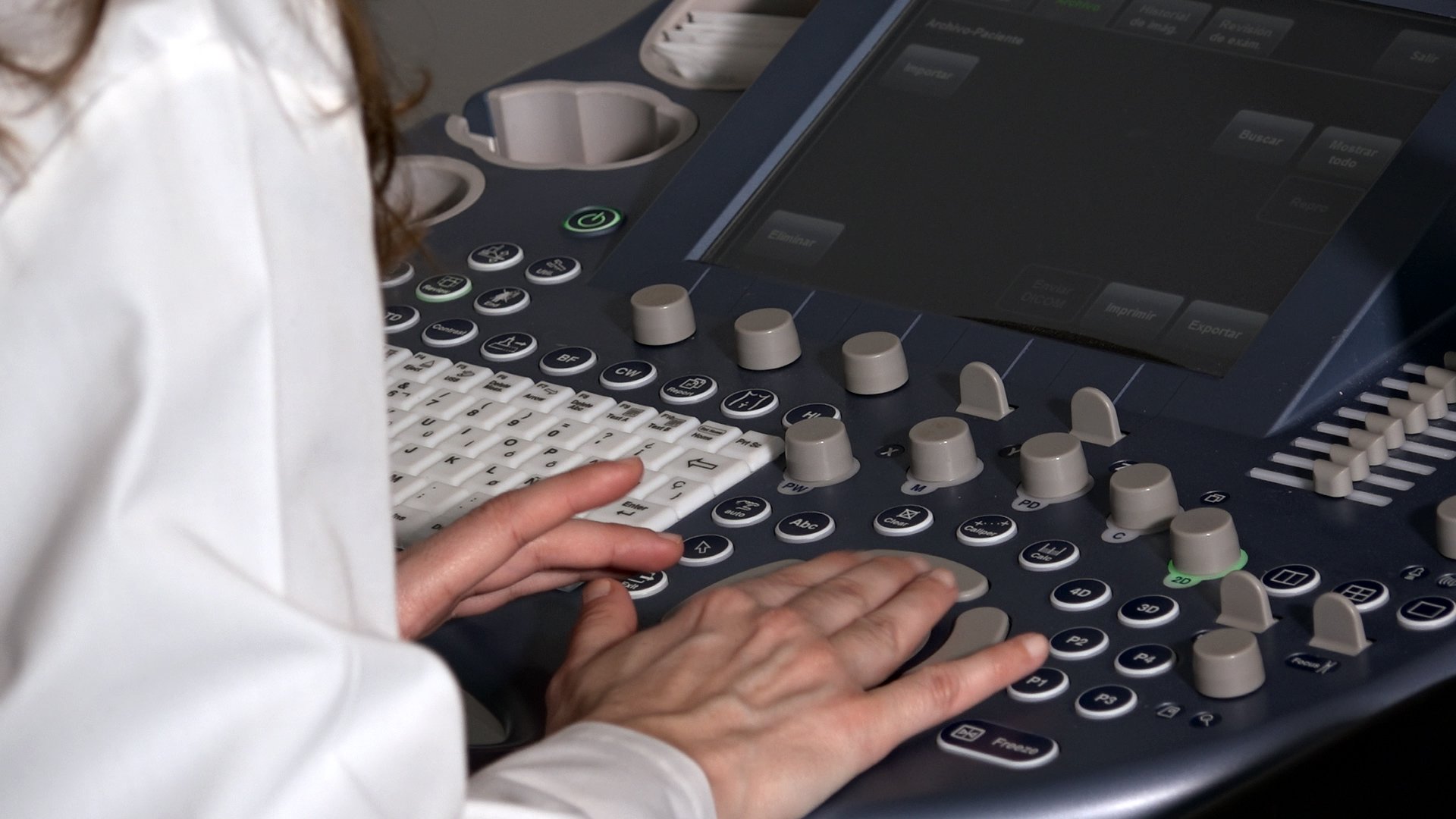
During the research work, which was carried out in its entirety at Instituto Bernabeu, 2D and 3D transvaginal ultrasounds were carried out on all patients due to undergo embryo transfer between 2014 and 2015. With the added extra of new HDlive high-definition or real-time US software, the reliability of the diagnosis is greater in comparison with standard findings when using conventional ultrasound techniques.
By processing coronal uterus images, in severe cases of adenomyosis, we get a more easily distinguishable 'tree formation' which is generally more easily recognisable in common adenomysis. It consists of an image which closely resembles a tree with the endometrium taking the form of the trunk and a clear representation of JZ rupture, perpendicular veins and hypoechoic masses which complete the branches and this is proportionately more variegated and dense in appearance, the more serious the case.
This is one of the 14 pieces of research work carried out at Instituto Bernabeu and accepted by the SEF scientific committee for its 31st National Congress to be held in Malaga between the 19th and 21st May.
A NEW ANDENOMYSIS ULTRASOUND MARKER F. Sellers, B. Moliner, J. Ll. Aparicio, A. Bernabeu, R. Bernabeu.
